
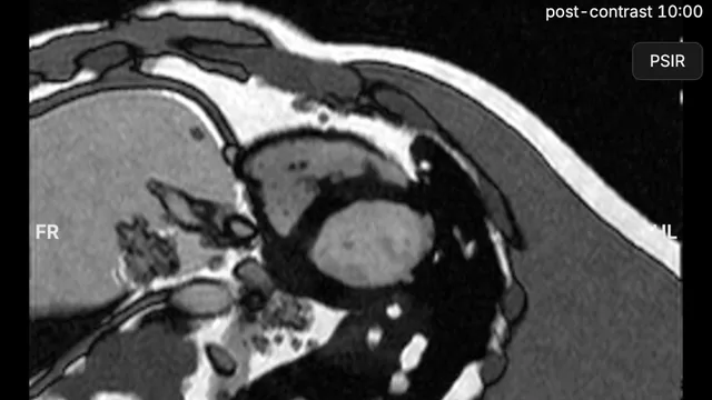
Short-axis cine stack planned on the 4-chamber and 2-chamber cines, mid breath-hold.
- 18clinical protocols
- Full studieslocaliser to late enhancement
- 1.5 T · 3 Tfield strengths
- Every stepgraded, with its correction
One platform for cardiac MRI training and education
Every acquisition is computed from the planned geometry and the sequence parameters on anatomy built from real volunteer scans, so the result of each decision shows in the next image.
The console follows a clinical scanner: program queue, planning panes, parameter card, physiology display and image viewer, on one screen.
- Virtual scannerComplete studies on a clinical-style console.
- Teaching modulesFrom MRI physics to every part of the exam.
- Graded assessmentEvery acquisition graded, with a report.
- Trainer dashboardProgress, courses and certificates for the whole class.
For you or your team
Your own licence
Radiographers, technologists, trainees and fellows can subscribe on their own, monthly or for a fixed term, with or without Assessment mode. Request a licence
Licences for your team
Named seats for departments, CMR courses and training programmes, with the trainer dashboard, courses and certificates under your institution's name. Request a quote

Planning
Planned on the images, as on the console
Slice groups are moved, rotated and resized on two reference images. Each view is built from the ones before it, from the pilots to the short axis.
- Field of view, phase direction, saturation band and adjustment volume shown on the plan
- Reference lines, package mode and perpendicular planning from the image
- In Learn mode, the ideal plane drawn for every step, with a live check
Read our guide: how to plan the cardiac MRI short-axis stack →
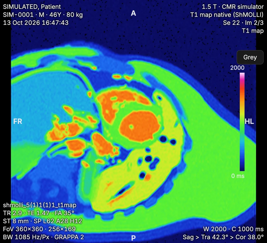
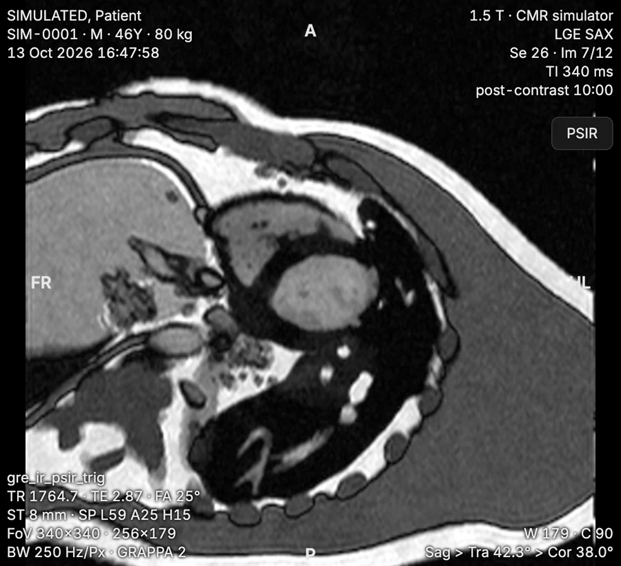
Image physics
Images that respond to every parameter
Signal, contrast and artefacts are calculated from the sequence and the anatomy, so MRI physics is learnt hands-on: a change on the parameter card shows in the next acquisition.
- Phase wrap, respiratory ghosting, off-resonance banding (worse at 3 T), Gibbs ringing, parallel-imaging aliasing, fat-water shift and implant artefact
- Cine, late enhancement with a TI scout, native and post-contrast T1 mapping with ECV, T2 and T2* mapping, perfusion and phase-contrast flow
- Scan time, voxel size, relative SNR and SAR recalculated with each change
Physiology
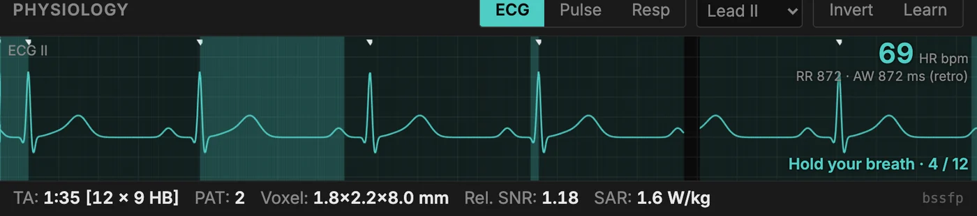
Physiology at real-scan timing
The ECG, breathing and contrast timing run at the pace of a real examination.
- ECG leads, trigger delay and acquisition window per beat, with arrhythmia rejection on retrospective gating
- Breath-hold commands in a recorded voice, and ghosting when a scan outlasts the breath-hold
- Patient conditions set at registration: heart rate, arrhythmia, atrial fibrillation, mistriggering, poor breath-holding and implants
Assessment
Every acquisition graded, every finding explained
Each step is checked against a written standard for plane, coverage, timing and image quality. The report shows the trainee, and the trainer, exactly what to practise next.
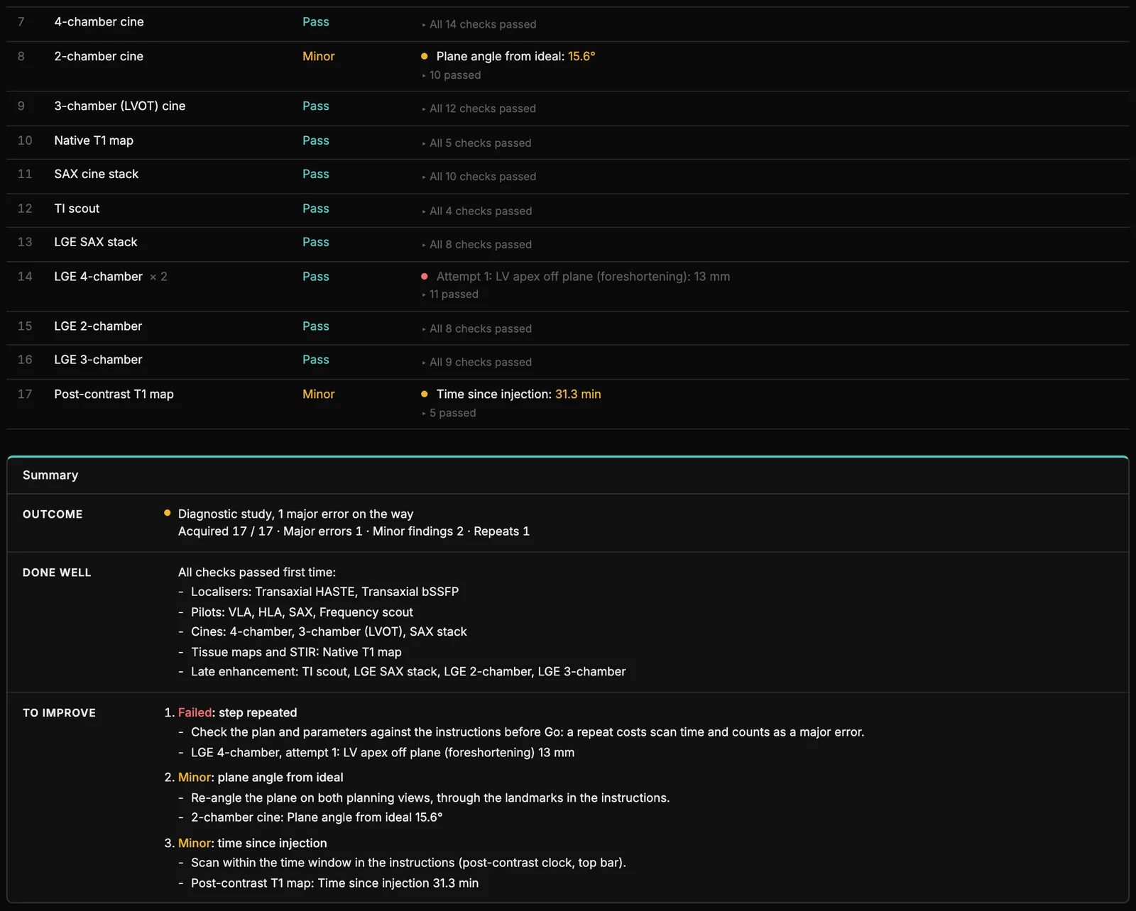
From an Assessment-mode exam: one step repeated after a foreshortened 4-chamber view, two minor findings, each with its correction.
- Each step and its resultPass, Minor or Critical, with the measurement behind each finding.
- An outcome for the whole studyDiagnostic or not, with major errors, minor findings and repeats counted.
- Specific correctionsEvery finding comes with what to change on the next attempt.
- What to practise nextA summary for the trainee, and a PDF mark sheet for the training record.
Training modes
From guided learning to independent acquisition
-
01
Learn
Instructions, a diagram of the ideal plane, a live plane check and Show me for every step, with the findings and their corrections after each scan.
-
02
Practice
The console with the step instructions only. Hints bring the full guidance back when needed.
-
03
Assessment
No guidance: the step names alone, with the simulated effects set by the trainer. The report grades the result and lists what to practise.
-
04
Clinical Scenarios
Referral-led examinations in which the patient changes during the scan: arrhythmia, devices, breath-holding and field strength to manage. Decisions are graded.
Teaching modules
Lessons from physics to planning
Interactive lessons, each ending on the console and with a certificate.
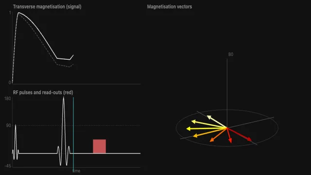
MRI physics: the basics
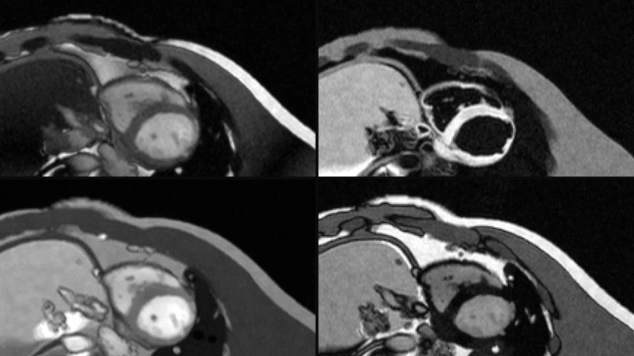
MRI sequences
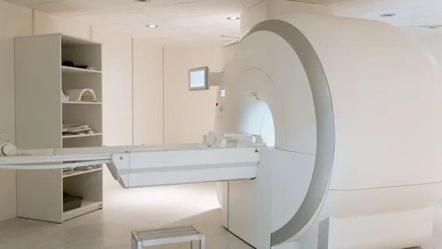
Safety checks
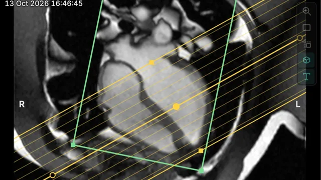
Cardiac views
Planning LGE
2D flow for valves
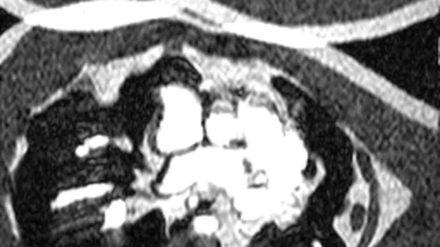
Which artefact is this?
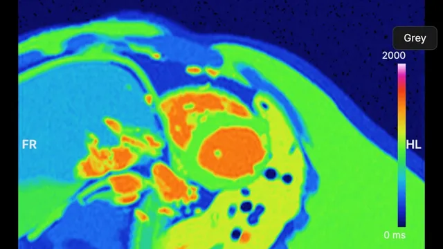
Mapping
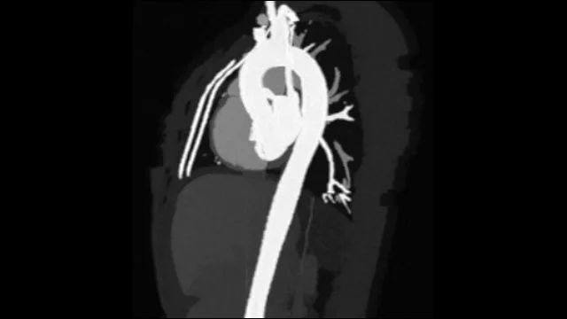
Contrast MRA
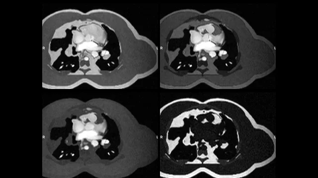
Dixon
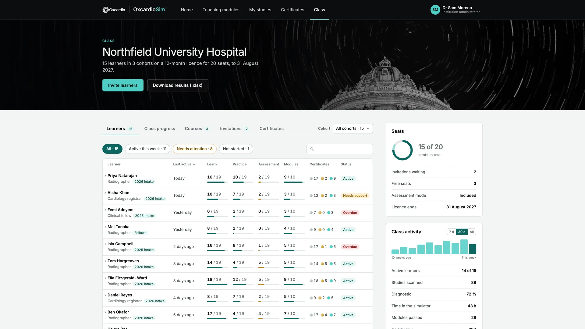
Trainer dashboard
Every learner's progress, in one place
For training leads: every learner's progress, courses and certificates, at a glance.
Viewing
Review the whole study
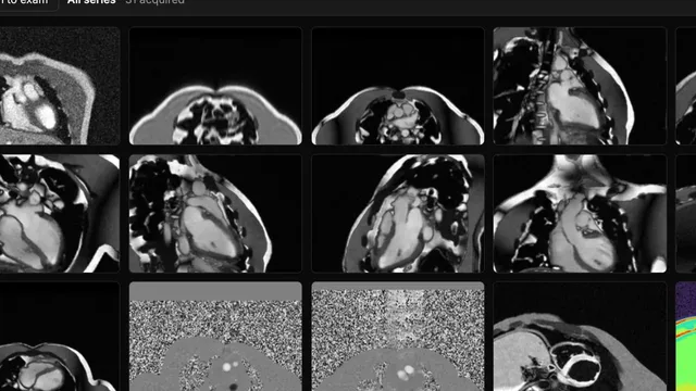
Every series of the exam in one gallery, from the localisers to post-contrast mapping, opened to any slice and frame.

Who it is for
MRI training for departments, courses and new services
Train without taking scanner time
New radiographers and technologists, clinical trainees and research fellows practise complete cardiac studies before and alongside supervised scanning.
Hands-on practice beyond the classroom
Course participants plan and acquire studies before and after the course, in their own time, with a graded report for each attempt.
Part of building a CMR service
Simulator access alongside our service development work: expert teaching, protocol optimisation and acquisition support.
Educational simulation, not clinical software. OxcardioSim produces simulated images for training. It does not process patient data, it is not a medical device, and it does not replace local protocols, safety procedures or supervised training on a scanner.
Questions
About the cardiac MRI simulator
What is OxcardioSim?
OxcardioSim is an MRI scanner simulator for cardiac MRI training. Trainees plan and acquire a complete CMR study on a simulated console. Each image is computed from the planned geometry and the sequence parameters, so every decision shows in the next acquisition.
Who is the simulator for?
MRI radiographers and technologists learning cardiac MRI or moving to a new scanner, cardiology and radiology trainees learning how the images they report are acquired, departments setting up or growing a CMR service, and educators running MRI courses.
What does it cover?
A complete cardiac MRI examination at 1.5 T and 3 T: localisers, planning of the long-axis and short-axis views, cines, phase-contrast flow, T1, T2 and T2* mapping, perfusion and late gadolinium enhancement with a TI scout.
ECG triggering, breath-holds and artefacts behave as they do on a scanner.
Which teaching modules are included?
Short guided modules sit beside the scanner: MRI physics and MRI sequences with live animations, MRI safety checks, the cardiac views, planning LGE, 2D flow for valves, T1, T2 and T2* mapping, contrast MRA, Dixon, and telling cardiac MRI artefacts apart. What is taught is then practised on the simulated console.
Do learners get a certificate?
Yes. Completed protocols and teaching modules earn a certificate, and departments can run courses that issue their own. Every certificate carries a code that anyone can check online.
A certificate records work done on the simulator; it is not an SCMR or EACVI level.
Is there a licence for a whole department?
Yes. Department licences give each learner a named account with Learn, Practice and Assessment, the teaching modules and Clinical Scenarios, plus an administrator account to manage learners, run courses and issue certificates. A pilot can come first.
Is it a general MRI simulator?
It is an MRI simulator built for cardiac MRI, including the aorta and contrast MRA. The MRI physics it teaches, from sequence parameters and image contrast to artefacts, scan time, SNR and SAR, applies to MRI of any region, so it also serves general MRI education. Neuro, musculoskeletal and abdominal examinations are not simulated.
How is training assessed?
Each step is checked against a written standard for plane, coverage and image quality, and graded Pass, Minor or Critical. Learn mode adds guidance and a correction for every finding. Assessment mode removes the guidance, and Clinical Scenarios also grade the decisions taken during the scan.
Is it tied to one scanner vendor?
The console follows the layout of a clinical scanner, and the planning and parameter decisions it teaches carry over to any vendor. Sequence names differ between vendors; the CMR-tool sequence guide maps them.
Can simulator training count towards CPD?
Radiographers and trainees can record simulator sessions as self-directed learning in their CPD portfolio.
Does it help where MRI scanner time is scarce?
Yes. Where scanners are few and fully booked, as in many hospitals across Africa, South Asia and parts of Southeast Asia and the Middle East, there is little room to train on patients. OxcardioSim gives radiographers and trainees MRI training on complete cardiac studies with no scanner time used, remotely and in their own time, and our acquisition training and protocol development can run alongside it.
Do we need to install anything?
No. OxcardioSim runs in a current web browser on a desktop or laptop, with no scanner time, patient or installation needed.
How does our department get access?
OxcardioSim is provided to partner institutions and training programmes. Departments and course providers can arrange a trial with their own team before committing. Get in touch to arrange one.