Home › Software › CMR planning guide

CMR planning guide

How to plan the cardiac MRI short-axis stack

The short-axis cine stack is the acquisition that LV volumes, ejection fraction and mass are measured from, so small planning errors carry straight into the report. This guide covers how to plan it, which settings to use and how to check the result before moving on.

At a glance

Planned on
The 4-chamber and 2-chamber long-axis cines (or the HLA and VLA pilots)
Orientation
Perpendicular to the LV long axis: the line from the apex to the centre of the mitral valve
Coverage
From the mitral valve plane at the base to beyond the apex
Slices
6 to 8 mm thick, with a 2 to 4 mm gap so slices are 10 mm apart
Sequence
bSSFP cine, temporal resolution 45 ms or better, one or two slices per breath-hold
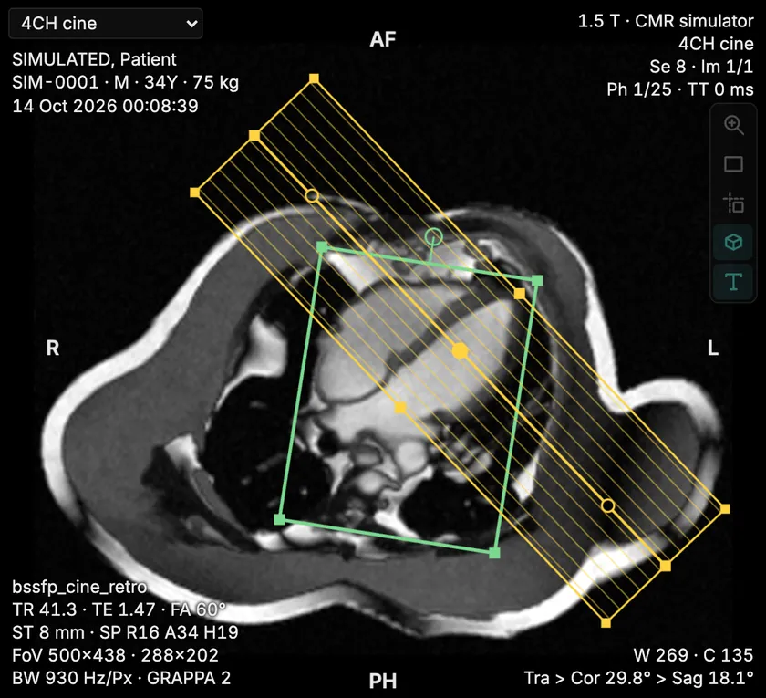
4-chamber cine with a stack of parallel short-axis slice lines perpendicular to the LV long axis, from the mitral valve plane to beyond the apex
Planned on the 4-chamber cine
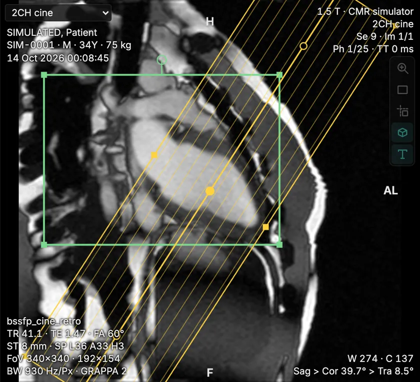
2-chamber cine with the same short-axis stack perpendicular to the line from the apex to the mitral valve
and on the 2-chamber cine
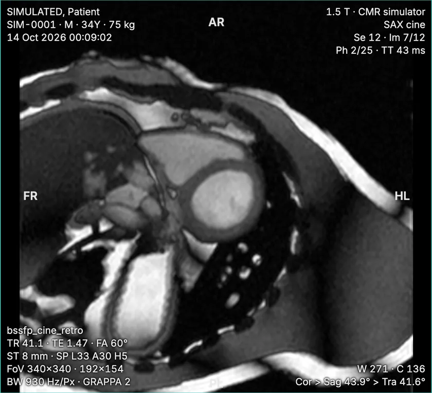
Mid-ventricular short-axis cine showing the round left ventricle and the right ventricle
Result: mid short-axis cine

Planning lines from OxcardioSim, our cardiac MRI simulator. Simulated images.

Why the short-axis stack matters

Volumes, ejection fraction and mass are calculated by contouring every slice of this stack at end-diastole and end-systole. The right ventricle can be measured from it too.

A stack that is tilted against the LV long axis cuts the myocardium obliquely, so the walls look thicker and the endocardial border blurs. A stack that stops short of the base or apex leaves volume out of the calculation. Neither can be corrected after the patient has left.

Before you plan: check the long axes

The stack is planned on the 4-chamber and 2-chamber cines, so it can only be as good as they are. Both should reach the true apex and pass through the centre of the mitral valve. If either is foreshortened, the long axis drawn on it is wrong and the whole stack inherits the tilt.

Planning on the HLA and VLA pilots instead is an accepted simplified approach, as long as the pilots themselves reach the apex.

How to plan it, step by step

  1. Load the 4-chamber cine in one planning pane and the 2-chamber cine in the other, at end-diastole, when the LV is largest.
  2. On the 4-chamber view, set the slices perpendicular to the LV long axis: the line from the apex to the centre of the mitral valve.
  3. Do the same on the 2-chamber view. Changing the angle in one view changes it in the other, so recheck both after every adjustment.
  4. Place the first slice at the mitral valve plane. Keep it perpendicular to the long axis rather than tilting it to lie along the valve: the SCMR protocol notes that the two are not always parallel.
  5. Extend the stack to just beyond the apex, so the last slice lies outside the LV cavity.
  6. Set slice thickness and gap: 6 to 8 mm slices with a 2 to 4 mm gap, so slices are 10 mm apart, and adjust the number of slices to the coverage.
  7. Check the field of view in the phase-encoding direction. Anything outside it, such as the arms or chest wall, wraps onto the image; enlarge the field of view or add phase oversampling if it would reach the heart.
  8. Coach the same breath-hold for every slice. Expiration gives the most consistent position; inspiration can be easier for some patients. What matters is that each breath-hold returns the heart to the same place.

Check the result before moving on

  • Coverage. On the long-axis views, the slice lines should span the LV from the mitral valve plane at end-diastole to beyond the apex. In systole the base moves towards the apex, so the most basal slice often shows atrium at end-systole: that is expected, and is why the end-systolic stack usually has one fewer basal LV slice.
  • Shape. The LV should look round in the mid-ventricular slices. An oval cavity means the stack is tilted against the long axis.
  • Breath-hold registration. Slices from different breath-holds should line up. A slice that appears out of sequence, or a border that jumps between neighbouring slices, points to a different breath-hold depth: repeat it.
  • Artefacts. Look for wrap from outside the field of view, and at 3 T for dark off-resonance bands across the heart on bSSFP. A frequency scout and a shim volume placed tightly over the heart move the bands away.
  • Rhythm. With ectopics or atrial fibrillation, use arrhythmia rejection with retrospective gating, or real-time cine if the rhythm or breath-holding prevents gated imaging.

Common errors

  • Planning on foreshortened long axes, so the stack is tilted from the start.
  • Angling the stack to follow the mitral valve instead of staying perpendicular to the LV long axis.
  • Stopping short of the base or the apex.
  • Uneven breath-holds, so slices are misregistered and volume is double-counted or missed.
  • Spacing slices more than 10 mm apart to save time.

Questions

How thick should short-axis cine slices be?

The SCMR standardized protocol recommends 6 to 8 mm slices, with or without a 2 to 4 mm gap, so that slices are 10 mm apart.

Should the short-axis stack be parallel to the mitral valve?

No. The stack is planned perpendicular to the long axis of the LV on the 4-chamber and 2-chamber views. The mitral valve plane is not always parallel to it, and tilting the stack to match the valve cuts the ventricle obliquely.

Where should the short-axis stack start and stop?

From the mitral valve plane at the base, at end-diastole, to just beyond the apex, so the whole LV cavity is included.

References

  1. Kramer CM, Barkhausen J, Bucciarelli-Ducci C, Flamm SD, Kim RJ, Nagel E. Standardized cardiovascular magnetic resonance imaging (CMR) protocols: 2020 update. J Cardiovasc Magn Reson. 2020;22:17. doi:10.1186/s12968-020-00607-1
  2. Herzog BA, Greenwood JP, Plein S, et al. Cardiovascular Magnetic Resonance Pocket Guide. 2nd ed. European Association of Cardiovascular Imaging (ESC); 2017.
  3. Herzog B, Kidambi A, Ballard G. CMR Congenital Heart Disease Pocket Guide. ESC Working Group on CMR; 2014.

Last reviewed: October 2026